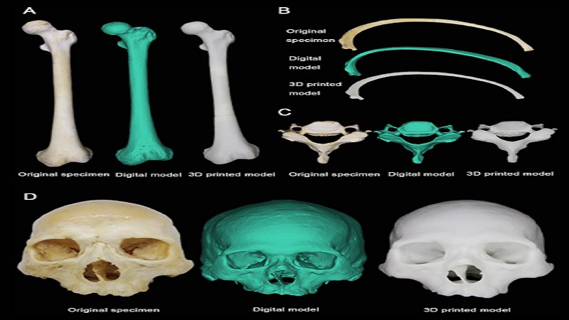
Researchers at Morphologic Science Experimental Center, Central South University, China, worked towards making the use of Phone Cameras and Cloud service-based workflow to image bone specimens and print their three-dimensional (3D) models for anatomical education. Using four typical human bone specimens, the femur, rib, cervical vertebra and skull , photographed by a phone camera, they aligned and converted them into digital images for incorporation into a digital model through the Get3D website and submitted to an online 3D printing platform to obtain the 3D Printed models. The results were excellent and as low as distance deviations ≤2 mm were noted among 99% of the random sampling points that were tested.
Read More: https://bmjopen.bmj.com/content/10/2/e034900.abstract
 Medical 3D Printing & Bioprinting